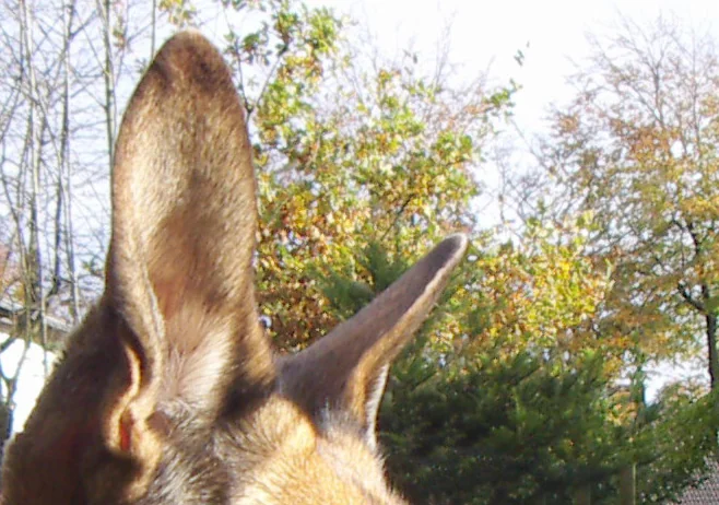
A close-up photograph of a dog’s ears, illustrating the outer-ear structures discussed in the article. Steffen Heinz (Caronna) · CC BY-SA 3.0 · Web size optimized; source linked for the original.
A dog’s ears do more than react to familiar household sounds. The visible outer flaps collect sound waves and help identify direction. Deeper structures then turn those waves into signals the brain can understand. Studies of canine ears describe the pinna, ear canal, middle-ear bones, and inner-ear cochlea in careful detail. This overview draws directly from veterinary research to show how these features support a dog’s sense of hearing. Any changes in a dog’s response to sound should be discussed with a veterinarian.
The Outer Ear and Sound Collection
The pinna is the visible ear flap. In many dogs it can rotate thanks to multiple muscles. This movement helps create small differences in the timing and strength of sound reaching each ear. Both upright ears and long pendulous ones direct sound waves into the ear canal.
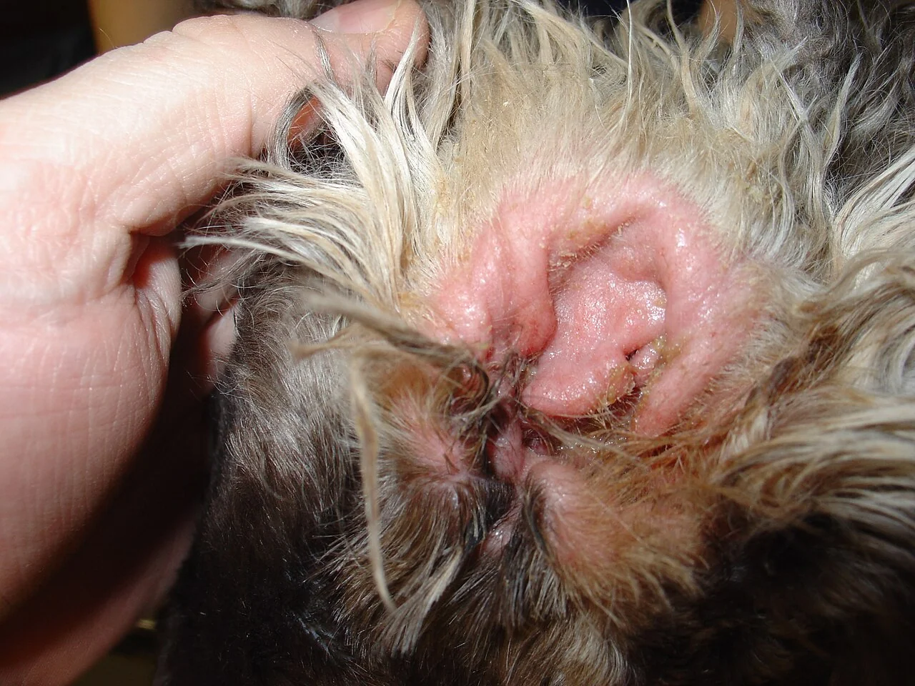
The ear canal is an L-shaped tube. Its length and angle differ by breed. The lining contains glands that produce cerumen. Observation of ear health forms part of routine care, but only under veterinary guidance.
Severe otitis externa in a four year old Cocker Spaniel showing the external ear structures. Joel Mills · CC BY-SA 3.0. Web size optimized; original linked.
The Middle Ear and Ossicles
Beyond the eardrum lies an air-filled middle-ear chamber. It holds three small bones: the malleus, incus, and stapes. These ossicles transmit vibrations from the eardrum toward the inner ear. Endoscopic views of canine heads confirm the presence of these structures along with the tympanic cavity and associated nerves.
The ossicles form a lever system. This arrangement helps move sound energy from air to fluid. Middle-ear cavity volumes and ossicular volumes vary with skull size across breeds, yet the overall shape of the ossicles remains similar. The middle ear also connects to the throat through the Eustachian tube, which equalizes pressure.
The Inner Ear, Cochlea, and Vestibular System
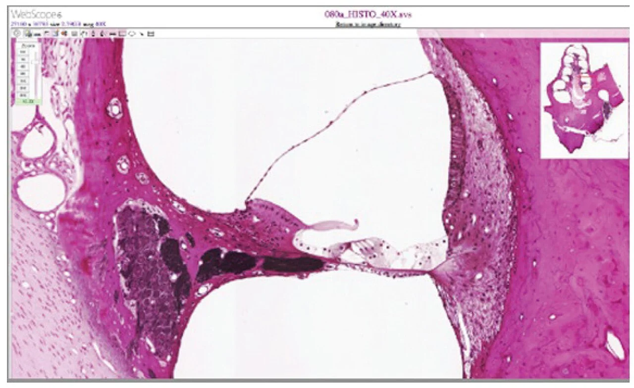
The inner ear sits inside the petrous temporal bone. The cochlea is a spiral structure. Inside it, the basilar membrane holds the spiral organ of Corti. Hair cells in this organ change mechanical waves into electrical signals sent along the auditory nerve.
The cochlea communicates with the middle ear through the round and oval windows. Nearby, the vestibular apparatus includes semicircular canals. These detect head movements and contribute to balance. Ear problems can sometimes affect steadiness.
Illustration of the cochlea from an anatomy and physiology textbook. OpenStax College · CC BY 3.0. Web size optimized; original linked.
Variation Across Breeds
Micro-CT scans of middle ears from 17 dog breeds plus a wolf show that cavity volumes, ossicular volumes, and related measures scale with negative allometry relative to skull size. A large breed’s middle-ear cavity can be many times larger than a small breed’s. Despite these absolute size differences, available behavioral hearing tests across breeds appear broadly similar.
The study notes that this similarity is unexpected under standard middle-ear models. It may reflect limits in current models or constraints elsewhere in the auditory system. Exact breed differences in everyday hearing remain an area of ongoing interest.
Everyday Observations and Veterinary Care
Dogs often move their ears when listening to nearby activity. The same structures that support detection of faint sounds also help interpret familiar cues. Understanding basic ear anatomy can make these moments more interesting to watch.
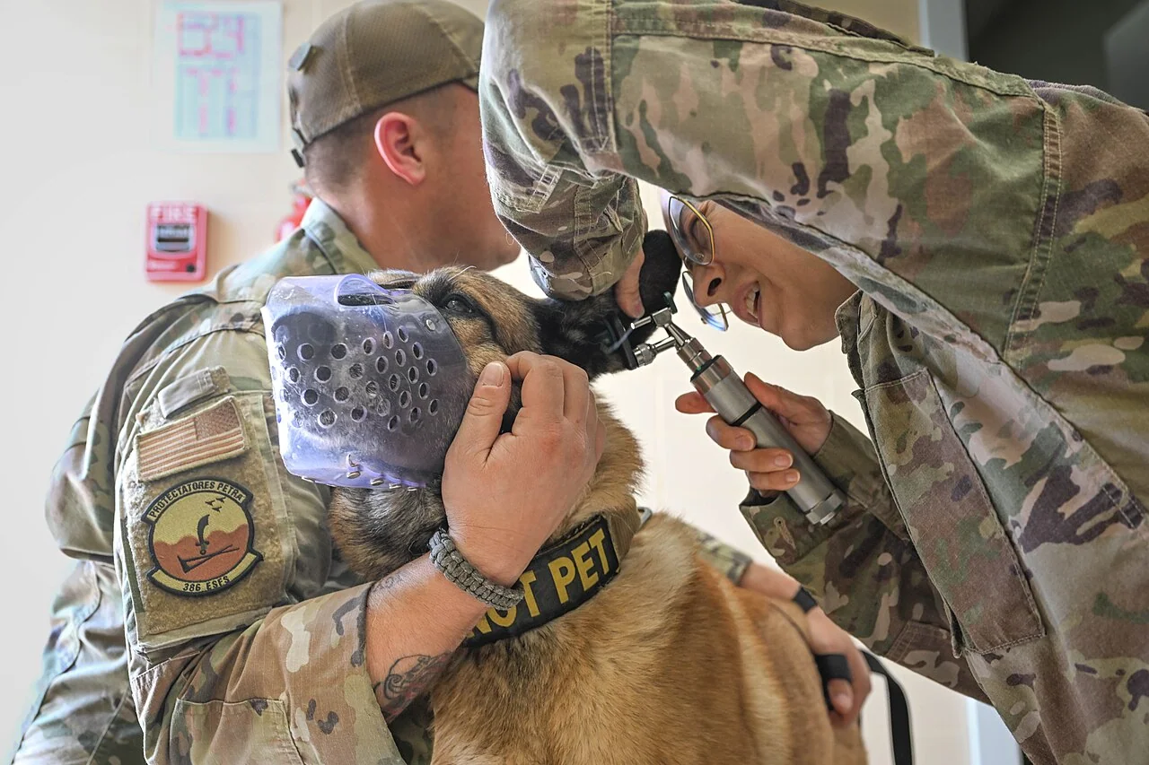
Changes in ear carriage or response to sound deserve professional attention. Veterinarians evaluate individual dogs using knowledge drawn from anatomical references. This article highlights structures described in peer-reviewed studies but does not replace clinical assessment.
General education, not individual veterinary advice. Ask a veterinarian about your dog’s health and care.
AI-assisted original writing. This feature was revised against source material and checked for unsupported claims. Follow the references below for evidence and its limits.
Research shows canine cognition follows a clear arc of early gains, midlife peak, and later gentle decline, offering owners practical ways to support their pets at every age.
Protected command centers in the brain and spinal cord team up with far-reaching nerves that connect to muscles, skin, and organs, letting dogs react to scents, sounds, and sudden surprises.
 Reference image