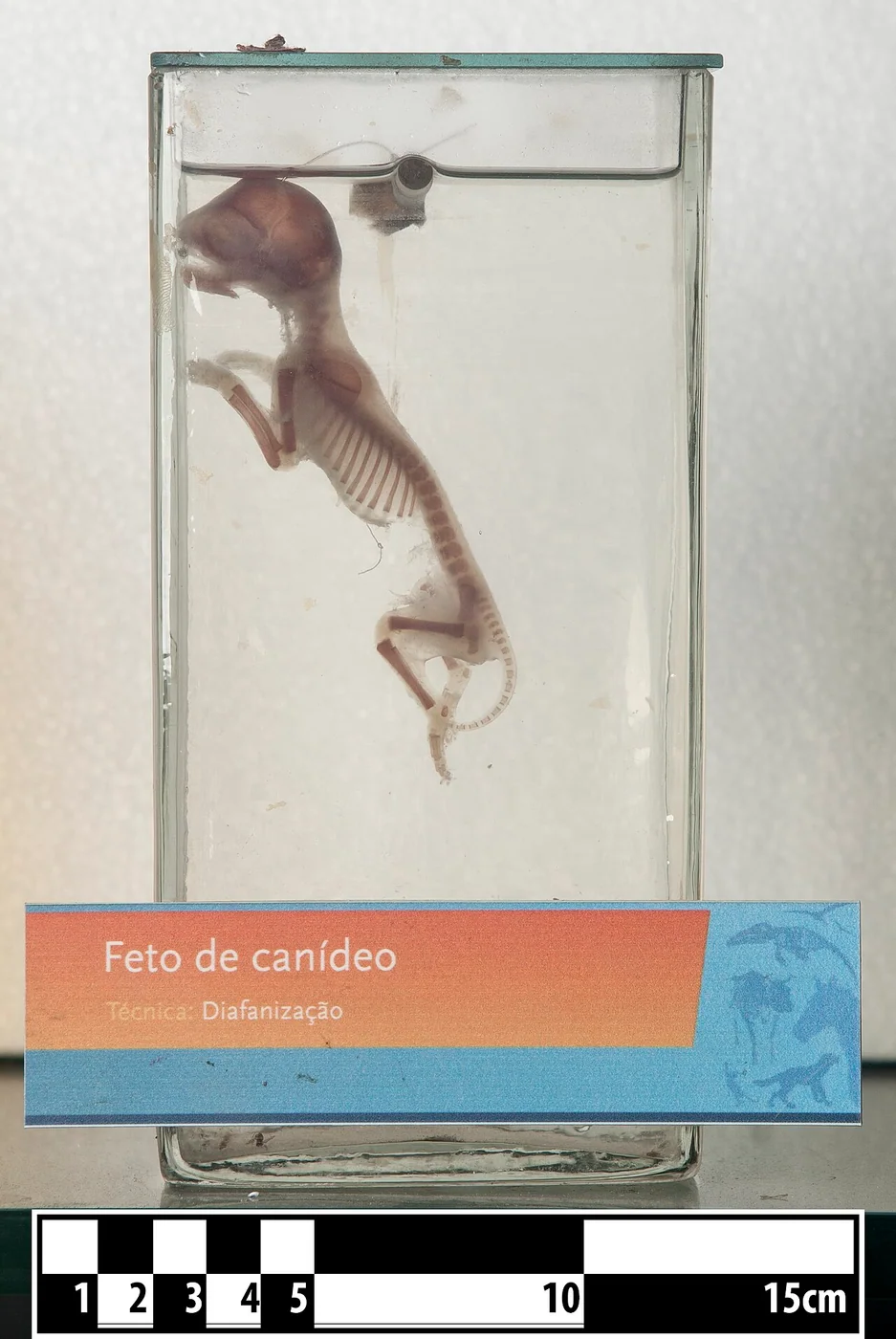
Protected command centers in the brain and spinal cord team up with far-reaching nerves that connect to muscles, skin, and organs, letting dogs react to scents, sounds, and sudden surprises.
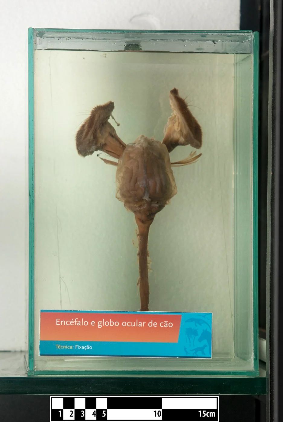
Dog brain and eyeballs, formalin-fixed anatomical specimen Source collection · CC BY-SA 4.0 · Web size optimized; source linked for the original.
A dog snaps its head toward a rustle in the grass or pulls its paw from a sharp stone. These quick moves depend on two working parts of the nervous system. The central nervous system, made up of the brain and spinal cord, processes information and sends commands. The peripheral nervous system carries signals between the central system and the rest of the body through cranial and spinal nerves. Both rely on neurons that connect to form circuits. Evidence shows the central parts sit inside bony protection and are wrapped in membranes called meninges filled with cushioning cerebrospinal fluid. Peripheral nerves travel outside this bony armor with thinner connective layers, making them more exposed to mechanical stress.
Central Structures and Their Safeguards
The central nervous system includes the brain, with its brainstem, cerebrum, and cerebellum, plus the spinal cord. The spinal cord is divided into cervical, thoracic, lumbar, sacral, and caudal segments that line up with regions of the vertebral column. Specialized tissues known as the meninges cover the brain and spinal cord. Cerebrospinal fluid surrounds these areas and helps protect them from everyday movement.
This setup lets the central system handle processing and coordination while staying shielded. The bony vertebrae and meninges reduce the effect of minor bumps that occur when dogs run, jump, or slide across floors.
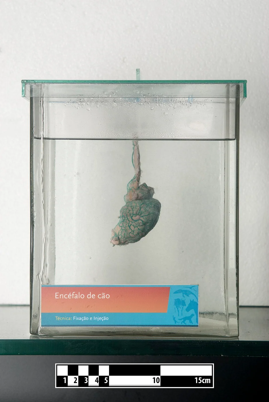
Dog encephalon, latex injection and formalin-fixed anatomical specimen Source collection · CC BY-SA 4.0. Web size optimized; original linked.
Peripheral Nerves and Their Reach
Peripheral nerves and ganglia sit outside the brain and spinal cord. They connect the central nervous system to the head, limbs, body, and internal organs. Pairs of spinal nerves and cranial nerves extend axons into the limbs, bladder, anus, and tail.
Sensory fibers in these nerves carry information from the skin and organs back to the spinal cord. Motor fibers travel outward from the central system to muscles. Dorsal roots bring mainly sensory axons into the spinal cord, with cell bodies in dorsal root ganglia. Ventral roots carry motor axons away from the spinal cord. These roots join to form mixed spinal nerves.
Because peripheral nerves lack the bony casing of the central nervous system, they face greater risk of mechanical injury from compression or stretching during normal activity.
Myelin and Signal Transmission
Both systems use myelin to speed electrical impulses along axons. In the central nervous system, oligodendrocytes produce this fatty coating. In peripheral nerves, Schwann cells form the myelin sheath. The changeover happens where the dura mater meets the perineurium at the start of spinal nerves.
Somatic lower motor neuron cell bodies remain in the central nervous system. Their axons extend through the peripheral nervous system to reach neuromuscular junctions. This division supports quick reflexes, such as pulling away from a painful touch, as well as longer coordinated movements.
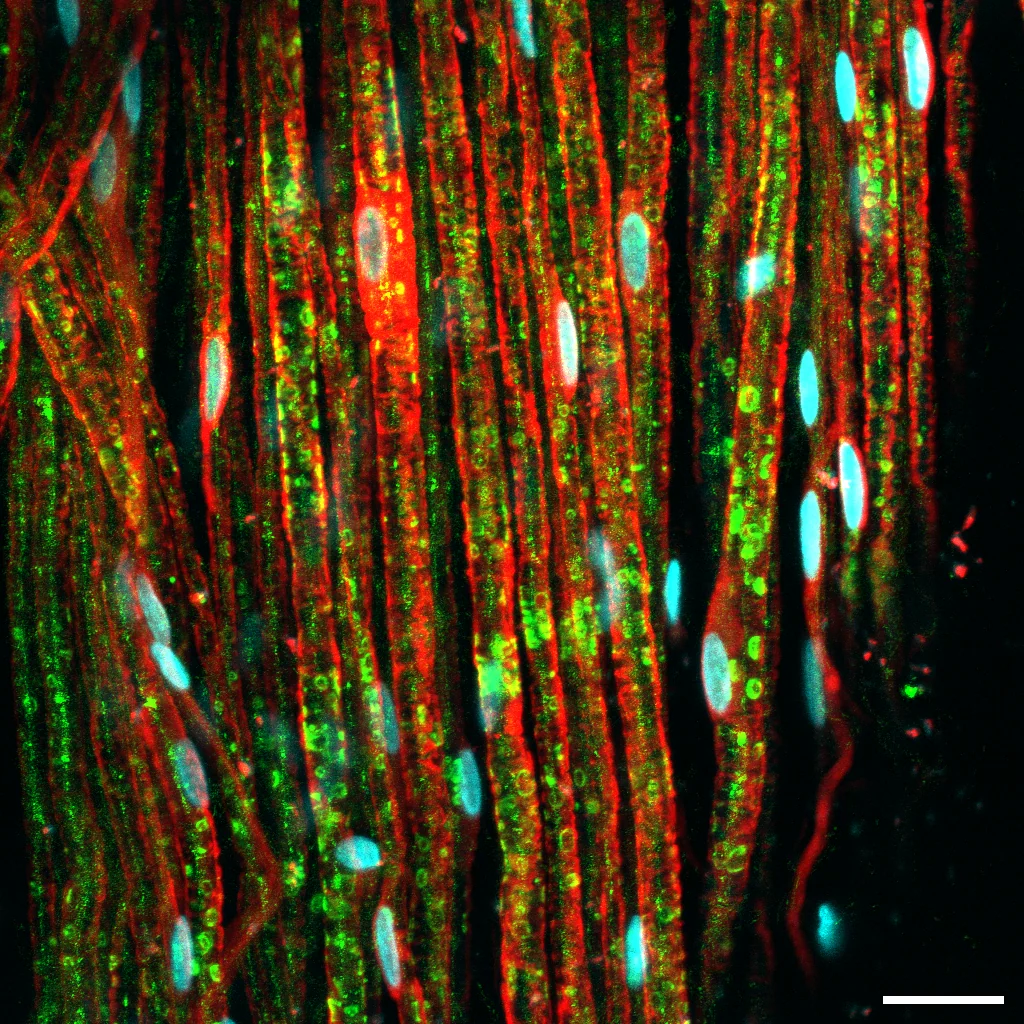
Immunofluorescence image of rat sciatic nerve showing myelin sheaths (green, MBP) and Schwann cells (red, S100). Valeriia Ustymenko · CC BY 4.0. Web size optimized; original linked.
Protective Layers Compared
Central nervous tissue benefits from the triple meningeal layers and the bath of cerebrospinal fluid that cushions and supplies nutrients. A selective blood-brain barrier further controls what substances reach the neurons.
Peripheral nerves rely on connective tissue sleeves called endoneurium, perineurium, and epineurium. These layers help nerves tolerate stretching during play or exercise but offer less defense against infection or trauma than the central coverings. The blood-nerve barrier in the periphery is also less restrictive, which can allow somewhat faster repair after minor damage.
Everyday Observations and Veterinary Advice
The contrast between protected central pathways and more exposed peripheral nerves appears in daily dog behavior. A sudden head turn toward a scent uses peripheral cranial nerves to carry the signal before central processing interprets it. A reflexive paw lift when stepping on something sharp can occur through spinal reflex loops before the brain registers the event.
Recognizing these differences reminds owners that changes in gait, alertness, or coordination should prompt a visit to a veterinarian rather than self-diagnosis. Regular checkups help catch shifts early in a system where small issues in one area can affect overall function.
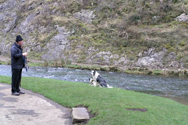
Border collies performing a paw-lifting obedience routine outdoors David Lally · CC BY-SA 2.0. Web size optimized; original linked.
General education, not individual veterinary advice. Ask a veterinarian about your dog’s health and care.
AI-assisted original writing. This feature was revised against source material and checked for unsupported claims. Follow the references below for evidence and its limits.
Research shows canine cognition follows a clear arc of early gains, midlife peak, and later gentle decline, offering owners practical ways to support their pets at every age.
 Reference image